
Nuclear stress testing is a non-invasive cardiac imaging procedure, and it helps diagnose conditions like coronary artery disease, heart failure, and arrhythmias. By evaluating blood flow to the heart at rest and during stress, the procedure gives cardiologists a detailed understanding of cardiac function. Here is more information on this testing procedure and what conditions it diagnoses:
What Is Nuclear Stress Testing?
Nuclear stress testing is a diagnostic imaging procedure used to assess how well blood flows through the heart muscle. A small amount of radioactive tracer is injected into the bloodstream, and a specialized gamma camera captures images of the heart at rest and under stress. These images help cardiologists identify areas of the heart that may not be receiving adequate blood supply, which allows for a more accurate assessment of cardiac function. The test is non-invasive.
What Conditions Does It Diagnose?
This type of testing is used to diagnose several cardiac conditions. Coronary artery disease (CAD) may be identified; the test reveals narrowed or blocked arteries by detecting regions of the heart with reduced blood flow during physical or pharmacological stress. Since blood flow is monitored at rest and peak stress, cardiologists can pinpoint the location and extent of arterial obstruction. Heart failure is also assessed. The imaging captures how efficiently the heart pumps blood with each beat, which allows cardiologists to calculate the ejection fraction, a key indicator of cardiac function.
Other diagnosable conditions include:
- Cardiomyopathy: a disease of the heart muscle that affects its ability to pump blood effectively
- Arrhythmias: irregular heart rhythms that may become more apparent under stress conditions
- Myocardial infarction (prior heart attack): areas of scar tissue appear as regions with no tracer uptake on imaging
What Does the Process Involve?
The procedure may take more than two hours, and it is conducted in two phases. These phases include a resting scan followed by a stress scan. During the resting phase, a radioactive tracer is injected, and images are taken to establish a baseline of blood flow, which allows the cardiologist to assess the heart’s condition at rest. Images captured during this phase serve as a reference point.
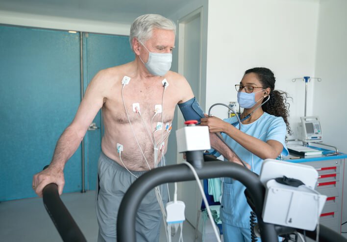
The stress phase follows the resting scan. The patient either walks on a treadmill or receives a medication that mimics physical exertion. Because the heart is placed under controlled stress, differences in blood flow become more detectable. A second set of images is captured during or shortly after this phase, and comparing the two sets allows the cardiologist to identify any reductions in blood flow that indicate underlying conditions.
What Are the Benefits?
Nuclear stress testing offers a level of diagnostic detail that standard stress tests cannot match, and this distinction is beneficial. The imaging component allows cardiologists to pinpoint the exact location and extent of reduced blood flow. Results are highly specific, so they support more precise diagnoses and targeted treatment plans.
The test is sensitive, and it can detect early-stage coronary artery disease before symptoms become severe. Early detection supports timely intervention. It provides data on how the heart is responding to ongoing treatment, which allows cardiologists to adjust care as needed.
Consult a Cardiologist Today
Nuclear stress testing is a diagnostic tool that provides cardiologists with a detailed understanding of cardiac health; it can identify coronary artery disease, heart failure, cardiomyopathy, and other conditions with a high degree of accuracy. The procedure is straightforward, non-invasive, and produces results that inform treatment decisions. If you have been referred for this test, speak with a cardiologist as soon as possible. Early diagnosis may lead to more effective treatment and better health outcomes.